Clinical Management of Delayed Corneal Re-Epithelialization Using Cryopreserved Amniotic Membrane
Introduction and Pathophysiology
Delayed corneal re-epithelialization following ocular trauma presents a complex clinical challenge. Normal epithelial defects should initiate closure within several days following baseline intervention. Failure to achieve prompt closure increases patient vulnerability to progressive complications, including stromal thinning, microbial keratitis, dense corneal scarring, and tectonic perforation.
Two principal clinical drivers underlie impaired epithelial wound healing:
- Neurotrophic Keratitis: Intact sensory innervation is essential for ocular surface homeostasis. Sensory nerve disruption compromises corneal metabolic and morphological pathways, suppressing normal cell turnover and migration.
- Corneal Exposure and Surface Desiccation: Incomplete eyelid closure, impaired blink dynamics, meibomian gland dysfunction (MGD), and delayed tear clearance exacerbate tear-film evaporation, provoking localized ocular surface inflammation.
Procedural Protocol for CAM Placement
Cryopreserved amniotic membrane (CAM) retains biologic growth factors that promote regenerative wound healing through anti-inflammatory, antifibrotic, and anti-angiogenic pathways. Application of self-retained CAM (such as Prokera Slim, BioTissue) yields accelerated epithelialization, superior visual outcomes, and fewer complications compared to bandage contact lenses alone.
| Procedural Stage | Clinical Action | Clinical Rationale |
|---|---|---|
| Preparation & Rinsing | Agitate CAM in an initial saline bath; transfer to a secondary sterile saline soak. | Removes storage media to minimize surface irritation. |
| Punctal Occlusion | Insert superior and inferior punctal plugs during soaking phase. | Preserves tear volume and elevates tear meniscus to maximize comfort. |
| Epithelial Debridement | Perform the “screwdriver test” across wound margins. | Identifies non-adherent, devitalized epithelium preventing migration. |
| Anesthesia Protocol | Avoid topical anesthetic drops during placement. | Ensures accurate, real-time patient feedback regarding ocular comfort. |
| Insertion Maneuver | Instruct downward gaze while tucking CAM under upper lid; direct upward gaze to position. | Self-seats membrane over the defect without abrasive mechanical friction. |
| Temporary Tarsorrhaphy | Apply Tarsus Eyelid Patch (Nictavi), folded cotton round, and adhesive amblyopia eye patch. | Immobilizes lid against the retaining ring, mitigating foreign-body awareness. |
[Mechanical Assessment: "Screwdriver Test"]
Perpendicular contact of Weck-cel sponge to corneal margin
                   │
                   ▼
Twist sponge gently against epithelial surface
       ├── Wrinkling Present  ──► Non-adherent; debride devitalized margin
       └── No Wrinkling       ──► Stable basement membrane; preserve margin
Patient Education and Postoperative Instructions
Patient expectation management mitigates anxiety surrounding foreign-body sensation and transient side effects.
| Domain | Patient Guidance |
|---|---|
| Expected Symptoms | Transient itching, burning, epiphora, ocular discharge, visual blurriness |
| Topical Therapy | Instill Optase Allegro (Scope) drops for localized pruritus |
| Activity Restrictions | Strictly avoid ocular rub and exposure to water or shower streams |
| Tarsorrhaphy Retention | Maintain protective patching to prevent lid sensation over the ring apparatus |
| Urgent Communication | Direct provider contact line provided for sudden or atypical pain |
Stepwise Escalation and Practice Management
Follow-up occurs after an initial 5-day retention interval. If the original CAM has dissolved and residual defect remains, a second CAM is positioned.
| Clinical Presentation | Advanced Therapeutic Intervention | Mechanism / Clinical Target |
|---|---|---|
| Recalcitrant Defect (Post-CAM 2) | Cenegermin-bkbj 0.002% (Oxervate, Dompé) | Recombinant human nerve growth factor for neurotrophic healing |
| Persistent Surface Fragility | Autologous blood serum or platelet-rich plasma (PRP) | Supplies neurotrophic and epitheliotropic growth factors |
| Meibomian Gland Obstruction | Fractional thermal treatment (Tixel-i, Novoxel) | Delivers thermo-mechanical energy to clear glands and stabilize tear film |
- Practice Workflow Optimization: Standardizing insertion on Wednesday and removal on Monday establishes predictable follow-up intervals for patients and clinical staff. Early intervention is indicated for persistent punctate epithelial erosions prior to progression to advanced ulceration.
Clinical Case Report
A female patient presented with an unhealed corneal epithelial defect refractory to 6 to 8 weeks of standard therapy and multiple bandage contact lenses.
- Intervention: Placement of a self-retained CAM was performed.
- Complication: The patient fractured the retaining membrane on the day of placement.
- Management: The CAM was disassembled from its rigid carrier ring, positioned directly into the inferior fornix, and secured via complete external tarsorrhaphy.
- Outcome: Evaluation 24 hours post-procedure demonstrated complete defect resolution with only a residual linear epithelial regeneration line visible.
Expert Quotations & Disclosures
“Epithelial defects should begin to heal within a few days of starting treatment. However, if a patient isn’t re-epithelializing as they should, it’s important to act quickly to prevent further progression, which can potentially lead to stromal thinning, infection, scarring or even perforation.”
“Before I place the CAM, my technician swirls it in a small glass of saline, then transfers it into a second glass of clean saline, where it soaks while I examine the patient and insert upper and lower punctal plugs for improved comfort (particularly in patients with low tear meniscus height).”
“I also do the ‘screwdriver test,’ where I touch the tip of a Weck-cel sponge perpendicularly to the epithelium and twist to see if the epithelium wrinkles; if it does, then it is loose and should be removed.”
“Even though this was far from an ideal placement, when the patient returned the next day, nothing was left but a regeneration line.”
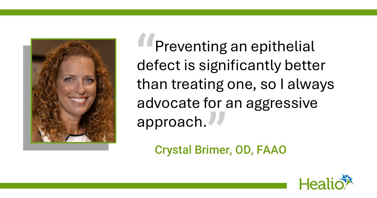
“Preventing an epithelial defect is significantly better than treating one, so I always advocate for an aggressive approach. If a patient has any level of persistent punctate epithelial erosion, I will place a CAM rather than waiting for it to reach stage 3 or 4 and potentially enter their line of sight.”
Author Contact: Crystal Brimer, OD, FAAO (crystalbrimerod@gmail.com)
Conflict of Interest Disclosures: Dr. Brimer reports personal fees and non-financial support from Bausch + Lomb, BioTissue, Harrow, Novoxel, Oculus, Optometric Aesthetic, Pure&Clean, and Scope.